
2.25million/year
Estimated annual volume of core microsurgical procedures in China: approximately 1.1545 million neurovascular, 512,100 malignant brain tumor related, 300,600 ENT, and 283,800 ophthalmic (excluding cataract).
DYNWAY
Microsurgical navigation for neurosurgery and microsurgery. It mounts onto the microscope already in the room and rests on two key innovations: sub-millimeter intraoperative real-time registration and micron-scale real-time instrument tracking.
Navigation has to keep answering “where am I”. Existing systems register once, before surgery, and fail the moment tissue deforms — continuous intraoperative registration remains unsolved.
The project remains in R&D and validation · Core technologies validated on the bench and in animal studies; end-to-end system navigation accuracy will be established by third-party testing after integrated prototype V1
PROJECT FILM
A full walkthrough of the operating principle, the prototype, and the experimental setup (with narration).
Clinical Need
The relative positions of lesion boundaries, instrument tips, and critical anatomy are difficult to judge directly. Critical boundaries and residual tissue are often below 0.5 mm — beyond the resolving limit of the human eye and the depth of field of an operating microscope — while conventional navigation accuracy of 1–3 mm sits orders of magnitude away from the 20–25 µm scale of microsurgery. As procedures move deeper and toward smaller structures, reliance on individual experience increases, along with training time and variability in risk.
Estimated annual volume of core microsurgical procedures in China: approximately 1.1545 million neurovascular, 512,100 malignant brain tumor related, 300,600 ENT, and 283,800 ophthalmic (excluding cataract).
Reported mean survival benefit of gross total versus subtotal resection for grade IV glioma.
Density is about 30% of that in developed countries, with a 10–15 year training cycle. In microvascular free-flap surgery, for example, novice anastomosis patency is only 60–70%, and roughly 600 cases are needed before the success-rate curve plateaus. Navigation supports decision-making and shortens the learning curve.
Procedure-volume data: China Health Statistics Yearbook. Other clinical figures are drawn from public research; definitions, reference years, and indication scope may vary between sources. The lack of deep-field and boundary information was confirmed in interviews with neurosurgeons at Sun Yat-sen University Cancer Center.
Limits of Existing Solutions
Conventional navigation is designed around rigid bony fixation, large reflective markers, and an open field. Once microsurgical resection begins, all three premises break down at once — drift, scale, and features compound, which is why intraoperative dynamic registration that follows soft-tissue deformation remains the key unsolved technical bottleneck.
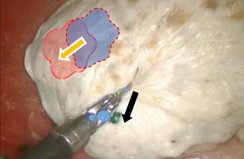
After the dura is opened, cortical shift can reach 1–2 cm (as reported in the literature), and every subsequent maneuver and resection step keeps changing tissue position. Existing navigation cannot re-register in real time, so the on-screen position gradually diverges from the actual surgical field.

Existing navigation carries system error of roughly 1–3 mm, insufficient for microscale boundary judgment. Conventional reflective spheres are about 11 mm in diameter against 0.3 mm microsurgical instrument tips, and readily obstruct a narrow field.

Soft tissue shifts non-rigidly as soon as the dura is opened, so the assumptions behind rigid-body and ICP-type registration no longer hold. A microscopic field spans only a few millimeters and offers very few stable, repeatable features, and glare, bleeding, and occlusion further alter the local image.
Sources: items 01 and 02 reflect feedback from neurosurgeons at Sun Yat-sen University Cancer Center; the magnitude of brain shift is from the literature (Nimsky et al. 2000; Nabavi et al. 2001); other figures are from manufacturers' published specifications and the literature; item 03 is a conclusion from the team's own algorithm validation.
Product Concept
Navigation rests on two conditions: where the instrument is right now and what the tissue in front of it is. Both must hold throughout resection. A system that answers them once, before the incision, is providing a plan — not navigation.
Objective: bring boundary, distance, and risk cues into the current view without disrupting the surgeon's core operating habits.Product value still requires continued validation in real clinical workflows, against defined baselines, and through surgeon feedback.
Key Technologies · Product Forms
The two key innovations are sub-millimeter intraoperative real-time registration and micron-scale real-time instrument tracking. In the product they take three forms: a split-path stereo operating microscope and trackable microsurgical instruments carry the instrument tracking, and the 3D navigation software carries the registration. The result appears as boundary and distance cues inside the surgeon's existing microscopic view. Together they form a layer of intraoperative perception.
Product Forms
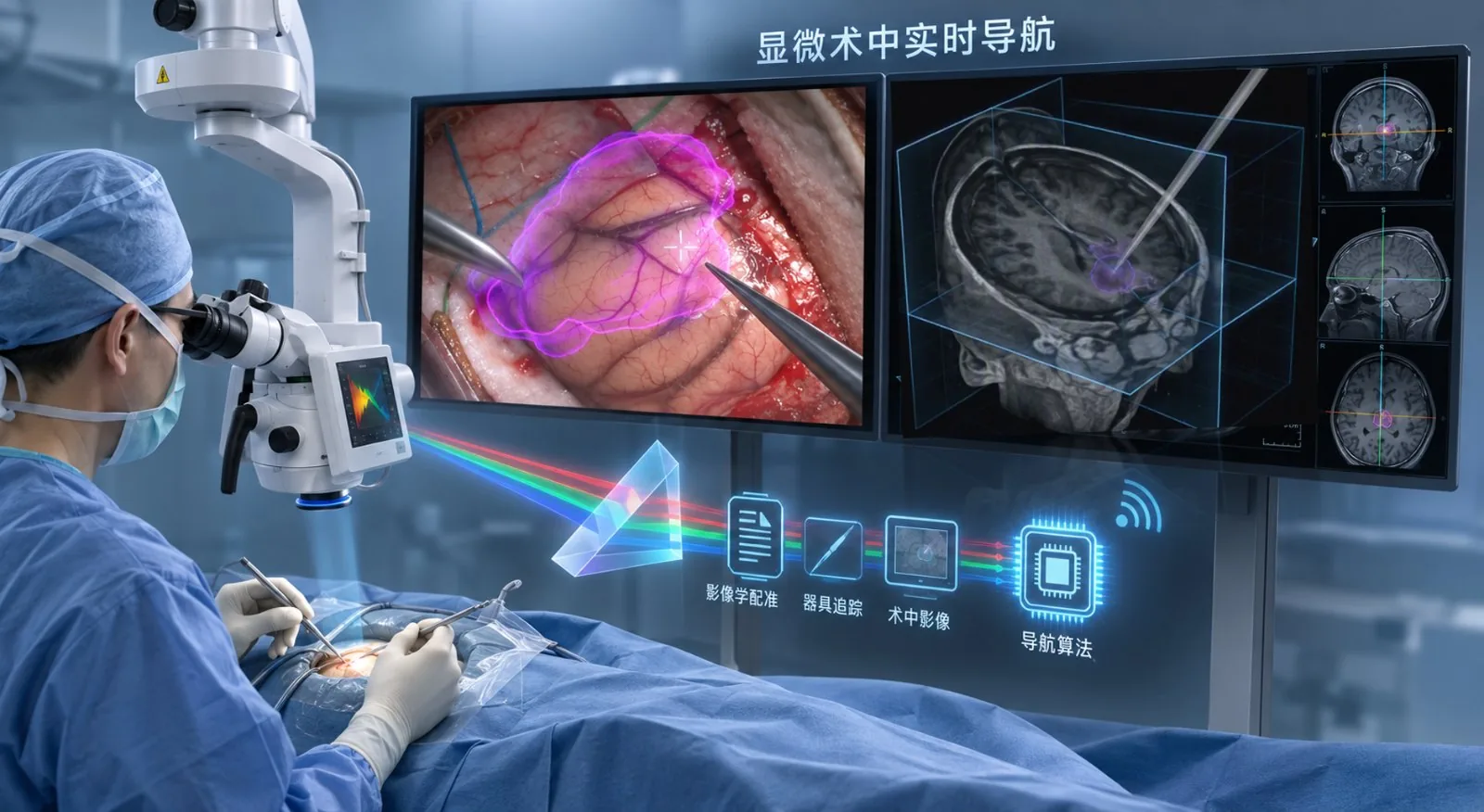
The system mounts onto the operating microscope already in the room and leaves the operating-room layout unchanged. The three forms marked here, plus the overlay inside the surgical field, are all of it that is visible in the operating room. The image is a concept rendering, not a photograph.
Select a numbered point in the scene, or any form below, to mark its position and open the technology behind it, with its study records.

01 · HARDWARE · SPLIT-PATH IMAGING
One optical path, two purposes. Coaxial beam splitting lets the same field capture visible and near-infrared light at once, with no filter-set switching: visible light renders the surgical field, and near-infrared picks up the signal from the fiber tracking points. The three images show the coaxial split path, the split-path module validation prototype, and integration with an operating microscope. The split-path module has completed bench validation.
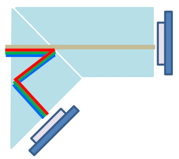


The microscope platform combines imaging and metrology, preserving the live surgical field while providing structural, depth, and relative-position references.
02 · INSTRUMENT · FIBER TRACKING
Optical fibers under 0.1 mm in diameter are embedded in forceps and scissors to form active tracking points at the tip, preserving the instrument's original dimensions, weight, and handling. Conventional reflective spheres are about 11 mm across, while a microsurgical instrument tip is on the 0.3 mm scale — so the tracking marker moves from a reflective sphere to a fiber at the tip, with tip position linked to the preoperative model.




The fiber-based active tracking point is covered by a patent application (Japanese Patent Application No. 2025-284200, accepted for filing), and proof of principle is complete; the precursor method, stereo keypoint 3D reconstruction, was published in IEEE T-ASE (2026).
03 · SOFTWARE · REGISTRATION
Registration runs in two steps. Initial registration uses semantic features such as vessel course and bone-surface texture in place of manually placed markers, needs no implanted bony markers, and has been validated in live-mouse craniotomies (ethics approval A2023M042-07, the University of Tokyo). Dynamic registration selects landmarks automatically on the tissue surface within the microscopic field, keeps assessing their reliability, and follows soft-tissue deformation; this step is currently validated on public data and in simulation, with in vivo validation scheduled for the next stage.
STEP 1 · INITIAL REGISTRATION · MEASURED IN LIVE-MOUSE CRANIOTOMIES
Semantic-feature constraints: vessel course and bone-surface texture replace manually placed markers, with no implanted bony markers. The table places three classes of registration method side by side and notes the source of each figure; soft-tissue deformation is handled by step 2, dynamic registration.
| Registration method | Registration error | Microscopic deformation | Source |
|---|---|---|---|
| Bone-anchored registration | approx. 0.8–2 mm | Not applicable | Published literature |
| Conventional ICP surface matching | 2.7–5.3 mm | Not applicable | Published literature |
| This project · semantic registration | 0.164 ± 0.059 mm (max 0.270 mm) | Handled by dynamic registration | Measured in live-mouse craniotomies |
The bone-anchored row is registration error; overall system error is typically 1–3 mm. Literature sources: intraoperative-CT registration, 0.86 mm; surface matching, up to 5.3 mm; ICP, 2.7 mm. Literature values and this project's measurements differ in experimental conditions, sample size, and statistical definitions and are not directly comparable; they are shown to indicate the difference in order of magnitude. This project's data come from live-mouse craniotomies, with a median translational registration error of 0.16 mm; the full statistical definitions and raw records are available in the diligence pack.


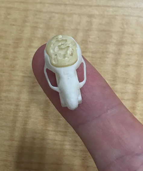

STEP 2 · DYNAMIC REGISTRATION · PUBLIC DATA AND SIMULATION
Dynamic landmarks are selected automatically on the tissue surface within the microscopic field, assessed continuously for reliability, and replaced as soon as they fail. Displacement of the deep lesion is inferred from the surface motion field, with no reference frame and no intraoperative imaging.
Selected automatically on the tissue surface, assessed continuously, and replaced on failure, keeping instrument trajectory, model, and live image in one spatial relationship.
After the view is abruptly rotated by 90° / 180°, tissue-tracking accuracy is 54.0% / 58.7%, against 4.6%–6.6% for existing point-tracking models under the same conditions. Dataset: STIR, a public dataset of in vivo soft-tissue surgical video. The test corresponds to recovering after the microscope is repositioned or the field is re-entered.
The lesion that is not directly visible is inferred from surface motion, with localization error 75% lower than without deformation compensation. Baseline: pose left unchanged after initial registration; organ simulation.
FAIL-SAFE
Bleeding, glare, and occlusion occur at any moment in the microscopic field. The system degrades in three steps according to registration confidence, and at no step does it keep issuing confident cues once the reference can no longer be trusted. The system advises and never acts; the decision always rests with the surgeon.
Outliers are rejected and the remaining landmarks maintain registration without interruption.
The overlay freezes at the last trusted pose and the system prompts for re-registration.
The overlay is withdrawn and the surgeon decides whether to re-register.
In vivo validation of soft-tissue deformation is scheduled for the next stage: ex vivo porcine tissue first, to test adverse conditions such as bleeding and glare, followed by 6–12 in vivo porcine studies within 2027, with results forming the animal-study evidence for the registration submission. The navigation-system patent, Japanese Patent Application No. 2025-284200, has been accepted for filing; precursor methods were published in Advanced Robotics (2024) and IEEE RA-L (2026).
These results demonstrate R&D-stage system capabilities. Medical use, performance claims, and clinical approval status remain subject to subsequent validation and public regulatory documents.
04 · INTRAOPERATIVE OUTPUT
The intraoperative output is how form 03 presents itself: boundary, instrument tip, and distance all appear inside the existing microscopic view, so the surgeon does not shift gaze. Real-time navigation is linked to the preoperative plan, visualizing the tumor boundary and flagging critical structures. The system advises and never acts.


Clinical Workflow
The system mounts onto the existing operating microscope. Staff positions, instruments, and screen layout stay unchanged; the only step added across the whole procedure is one registration after the microscope is positioned. The surgeon receives boundary and distance cues inside the original microscopic view without shifting gaze.
Import MRI/CT, segment the lesion and critical structures, and plan the approach.
Performed once after the microscope is positioned, with no implanted bony markers. This is the only step the system adds.
The system continuously evaluates and updates the correspondence between the preoperative model and the live field, with no surgeon input.
Presents lesion boundary, instrument tip, and relative position to critical structures inside the microscopic view.
Retains process data for case review, training, and accumulation of clinical evidence.
Sterility and instrument handling follow existing practice: the split-path module is covered with a single-use sterile drape without changing draping procedure, and fiber instruments are sterilized as conventional microsurgical instruments. Scrub staff add only drape placement and counting. Actual steps and per-stage timing are still being confirmed with collaborating clinicians.
Development and Intellectual Property
Proof of concept is complete, patents have been filed and granted, and seed-round investment commitments are in place. The product path runs through module prototypes, integrated prototypes, registration testing, clinical trials, and NMPA review. Timing remains dependent on experimental progress, regulatory consultation, and funding.
Completed method innovation and the core technical route, advanced human-machine interaction validation and first-generation prototype development.
Animal studies and validation of the split-path camera module prototype completed; engineering iteration of the navigation software and instrument-tracking system under way; the company, Dynway Technology Co., Ltd., was established this year.
Two integrated V1 prototypes; registration testing starts in Q3, and pre-submission consultation with the review authority in Q4 fixes the clinical evaluation pathway.
A supplementary small-sample clinical trial, alongside registration prototypes and quality-system audit preparation; research-grade modules and OEM collaboration bring first revenue.
Registration documentation submitted, entering technical review; Q3 2028 at the earliest.
Includes buffer for review deficiency responses. This target depends on regulatory consultation and trial progress; the final timeline will reflect actual development.
Commercialization does not hinge on this milestone: the Training & Assessment mode, OEM modules, and software licences generate revenue before approval. The staged path is set out in the Market, Competition, Funding and Milestones section below.
The team has 73 papers in this field, 15 of them core papers (including in IEEE T-ASE and RA-L) and 11 with a team member as first author; the robotic autonomous mouse-craniotomy drilling system received the IROS 2023 Best Application Paper Award. 74 animal studies have been run to date under ethics approval. The research collaboration network spans four institutions in China and Japan: Sun Yat-sen University Cancer Center (Neurosurgery) and the University of Tokyo Faculty of Medicine (ophthalmology, pediatric surgery) have signed letters of intent for clinical validation; Zhongda Hospital, Southeast University, and Teikyo University School of Medicine have expressed intent for research collaboration.
Electrical safety, electromagnetic compatibility, software, and biological evaluation, against GB 9706.1, YY 9706.102, GB/T 25000.51, and GB/T 16886; testing is planned at a provincial medical-device testing institute.
Pre-submission consultation with the review authority in Q4 2027 fixes the pathway, followed by a supplementary small-sample clinical trial with ethics approval and filing. Marketed surgical-navigation products serve as the comparable devices; because the core principle differs, the baseline plan is a single-arm trial with an accuracy endpoint at 2–3 centers.
Registration units and submission documents; manufacturing licence, quality-system audit, and supply-chain and process freeze. Two routes: contract manufacturing (asset-light, allows earlier acceptance) or an in-house assembly and test line; contract manufacturing is preferred.
Registration documentation submitted, entering technical review; Q3 2028 at the earliest. The statutory technical review is 90 working days, plus quality-system inspection and administrative approval; with deficiency responses the practical estimate is 12–24 months.
One deficiency round is budgeted at 3–6 months and is already included in the 2030 approval target; quality-system documentation is prepared in parallel and the pre-submission consultation starts early. These dates are planning figures; the final timeline depends on regulatory consultation and trial progress.
These are R&D-stage results from bench and live-mouse measurements. Raw calibration and measurement records are available in the diligence pack; end-to-end system navigation accuracy will be established by third-party testing after integrated prototype V1.

Study details are in the preprint arXiv:2609.12530 (submitted to IEEE T-RO).
| Year | Paper topic | Journal / conference | Team authors | Relates to |
|---|---|---|---|---|
| 2026 | Bone micro-milling with automatic calibration and 3D surface fitting | IEEE RA-L | Enduo Zhao (first author), Xiaofeng Lin | Calibration and registration · ex vivo mouse 6/7 |
| 2026 | Microsurgical needle handling with real-time stereo keypoint tracking | IEEE T-ASE | Xiaofeng Lin | Instrument tracking (the object is a suture needle) |
| 2025 | Autonomous drilling of mouse cranial windows | IEEE T-ASE | Enduo Zhao (first author) | Validation platform · ex vivo mouse 14/20 |
| 2025 | Estimating the state of the drilled object through active contact | IEEE RA-L | Xiaofeng Lin (first author), Enduo Zhao | Validation platform |
| 2025 | Completion recognition in autonomous micro-drilling | Scientific Reports | Enduo Zhao (first author) | Validation platform |
| 2025 | A multimodal digital twin for micro-drilling | IJCARS | Enduo Zhao | Validation platform |
| 2024 | Skull-feature registration with a microscopic stereo camera | Advanced Robotics | Xiaofeng Lin (first author) | Registration · precursor method |
| 2023 | Autonomous drilling of mouse cranial windows: evaluation on an egg model | IROS · Best Application Paper Award | Enduo Zhao (first author) | Validation platform |
| 2026 | Precision milling with general anatomical priors and active boundary sensing | Preprint arXiv:2609.12530 · submitted to IEEE T-RO | Enduo Zhao and Xiaofeng Lin (co-first authors), Yuhan Song | Registration 0.16 mm · live mouse 15/15 |
The first eight are peer-reviewed publications and the last row is a preprint; the manuscript on dynamic landmarks is not yet published and is not listed. The “paper topic” column summarizes each paper and is not its title. The team has 73 papers in this field, 15 of them core papers and 11 with a team member as first author; DOIs can be checked in the diligence pack.


IP PORTFOLIO
Counted by patent family (the Chinese and WIPO force-feedback filings count as one). The two patents that map directly onto the current product are the core navigation applications, both accepted for filing: the Japanese application for a navigation system, algorithm, and instruments (Japanese Patent Application No. 2025-284200) and the Chinese application for a 3D calibration board and performance-evaluation method for stereo microscopic imaging (CN202610805057.1). The six granted patents are in surgical robotics and reflect prior technical work; all eight name CEO Xiaofeng Lin as inventor. The barrier is structural: sub-millimeter intraoperative real-time registration × micron-scale real-time instrument tracking, combined in one hardware-software system. Ownership, licensing scope, and freedom to operate require item-by-item confirmation during investment and partnership due diligence.
Team
R&D, algorithms and AI, clinical work, technology transfer, business and supply chain, company and capital — each core step in taking a device from the lab to the hospital has an owner. Core members have research and academic experience at the University of Tokyo, Tsinghua University, Harvard University, Yale University, the University of Chinese Academy of Sciences, and Harbin Institute of Technology; two advisors cover clinical translation and industry and registration.

FOUNDER & CEO
Project Researcher in medical devices, Faculty of Medicine, the University of Tokyo · Leads product, technology, research and clinical work, and strategy
 Guanhao Lin
COO
 Enduo Zhao
CTO
 Yuhan Song
R&D Director
 Zenghui Yu
Business Director
 Shenghao Jiang
Clinical Research
 Gina Quan
Tech Transfer
 Jian Lu
Clinical Advisor
 Chusheng Wu
Industry Advisor
COO · Co-founder
CTO · Co-founder
R&D Director · Co-founder
Business Director · Co-founder
Clinical Researcher
Technology Transfer Researcher
CLINICAL TRANSLATION ADVISOR
Supports: the technology-transfer pathway; access to resources at Southeast University and Zhongda Hospital; clinical application and extension across departments
INDUSTRY ADVISOR
Supports: access to industry and supply-chain resources; guidance on the quality system and registration preparation; industry strategy
Market · Competition · Funding & Milestones
The project remains in R&D and validation. One system runs in two modes: before approval, revenue starts through the Training & Assessment mode, OEM modules, and software licences, so commercialization need not wait for full-system approval; after approval the same system switches to Surgical Navigation mode and the business moves to proprietary systems, software, and consumables.
01 · Position
Navigation has to keep answering two questions throughout resection: where is the instrument and what tissue is this. Existing products each have their strengths: some already bring the navigation image into the microscope, and some achieve continuous registration on exposed bone. What this product does differently is to do three things at once — cover the field under the microscope, track the instrument itself inside the microscopic view, and keep registering during surgery as tissue deforms.
| Product / country | ①Extends to microsurgery | ②Instrument tracking in the microscopic view | ③Continuous intraoperative registration | How it registers |
|---|---|---|---|---|
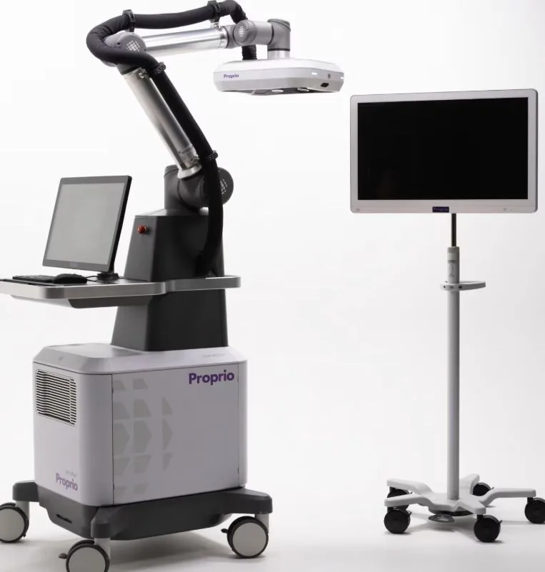 Proprio ParadigmUnited States |
No | No | Yes | Continuous registration on rigid anatomyA camera array matches exposed bone to the CT model in real time, registering each vertebra rigidly; designed for open spine surgery. |
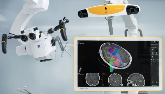 Brainlab Microscope NavigationGermany · on Zeiss / Leica microscopes |
Yes | No | No | Tracks the pose of the microscopeTracks microscope pose and overlays preoperative images in the eyepiece; the overlay comes from the single preoperative registration. |
 Medtronic StealthStation S8United States |
Yes | No | No | One registration, updated by intraoperative rescanInfrared / electromagnetic tracking of a reference frame, with the navigation image injectable into the microscope eyepiece; registration is updated by rescanning during surgery. |
 Beyeonics OneIsrael · digital microscope |
N/A | No | No | A digital microscope, positioned as visualizationThe surgical field is digitized and sent to a head-mounted display; it includes no image registration or navigation function. |
 7D FLASHUnited States |
No | No | No | Registration in seconds, one snapshot at a timeA reference frame clamps the vertebra and visible light matches exposed bone; after the anatomy changes, a new snapshot updates it. |
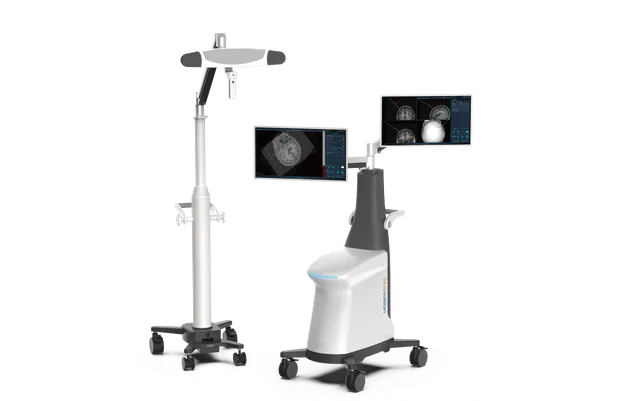 Huake PrecisionChina · neurosurgical robotics |
No | No | No | One registration before the craniotomyStructured light scans the surface of the head and face to register; registration is done before the craniotomy and carried through after it. |
 Dynway intelligent navigation for microsurgeryThis product · meets all three within this table |
Yes | Yes | Yes | Continuous registration that updates with tissue deformationRegisters to the tissue itself inside the microscopic view; no reference frame and no intraoperative rescan. Overall system accuracy and clinical value are still being validated. |
Criteria: ① takes conventional (non-microscopic) surgical navigation as the baseline and asks whether the product goes on to cover the field under the microscope; Beyeonics One has no navigation function, so it is marked not applicable. ② means image-level tracking of the instrument itself inside the microscopic view, excluding tracking of external optical markers. ③ means registration is updated continuously and automatically during surgery from the anatomy itself, without rescanning; tracking only a rigid reference frame does not count. This product's intended specialties are neurosurgery, ENT, ophthalmology, and reconstructive surgery.
Compiled in November 2026 from each manufacturer’s public materials and registration information. Competitor specifications and approval status change over time; current specifications, indications, and registration status are subject to each manufacturer’s own disclosures. Product names and trademarks belong to their respective owners and are used here for identification only.
02 · Market → 03 · Revenue
Neurovascular, malignant brain tumor related, ENT, and non-cataract ophthalmic procedures combined.
Boundary judgment directly determines the extent of resection and survival benefit, the clearest value proposition.
A target pool of about 120 leading neurosurgical centers; then ENT, ophthalmology, and reconstructive surgery.
Same hardware, with software enabled by mode. Education and research only, not for diagnostic or treatment decisions; for microsurgical skills centers at medical schools and teaching hospitals, microsurgery training centers, and animal laboratories. Training customers become the first installation candidates.
Hardware module delivery; software licences bundled with hardware and billed annually; engineering services for microscope OEMs only, one-off per model. Customers start with university and research-institute laboratories, then microscope manufacturers and navigation-system vendors (including surgical-robot makers), validating demand, interfaces, and willingness to pay. Tertiary neurosurgery centers are the installation target after approval.
The same system switches to Surgical Navigation mode. Complete-system installations build scale; enhanced software licences renew annually; dedicated consumables launch after approval, forming recurring revenue.
One set of hardware and one registration and tracking stack, with software enabled and charged by mode. Before approval, experience is turned into measurable indicators and revenue; after approval, the system enters the operating room. Commercialization and registration run in parallel, releasing risk in two steps.
Sources: procedure volumes from the China Health Statistics Yearbook; market size from a published market report by Grand View Research. Third-party forecasts rest on their own definitions and assumptions and may differ from actual outcomes.
04 · Risks, Funding and Milestones
The next priority is converting technical performance into clinical evidence and a registrable product. The innovation is concentrated in perception and registration; the system advises and never acts, and the decision always rests with the surgeon. Each of the three risks has a matching action, and exposure is released in stages.
Seed-round investment commitments are in place from several individual investors, funding 18 months of work and carrying the company to Pre-A; prizes and government grants serve as non-dilutive supplements. The path runs from closing to prototype delivery, third-party validation, hospital proof-of-concept, and the start of Pre-A, with each deliverable tied to a disbursement point.
Counted from the closing date; IP work begins and the Guangzhou base and operating foundation are set up.
Two units suitable for demonstration and third-party testing; the microscope interface specification is finalized.
Phantom accuracy, biocompatibility, and electromagnetic-compatibility testing; classification consultation.
Target: proof-of-concept agreements with at least two hospitals and interface-validation talks with one OEM; Pre-A begins.
RESOURCE PRIORITIES
MILESTONES
The basis and sources for procedure volumes are given in the Clinical Problem section above. Market size and commercial path illustrate the commercial logic; actual performance will depend on installation ramp, gross margin, channel discounts, and the post-registration launch schedule. Milestones are planning figures; actual dates depend on the closing date and validation progress. Specific pricing, installation plans, the round size, and detailed use of funds are calculated against the milestone budget, cash flow, and registration plan, and are not disclosed on this page. They are available in formal due-diligence materials.
CONTACT
We welcome direct contact from neurosurgical and microsurgical teams, microscope and instrument manufacturers, and investors following early-stage medical device programs.
Full due-diligence materials — covering the clinical validation plan, registration pathway detail, financial projections, and use of funds — are available on request. Please state your organization and intended use.
Enable JavaScript to view the email address WeChat · scan or long-press the code
DYNWAY
Dynway's first product is an intelligent navigation system for microsurgery, built on two key innovations: sub-millimeter intraoperative real-time registration and micron-scale real-time instrument tracking. Together they form a layer of intraoperative perception — the foundation of the perception layer for embodied surgical intelligence, and what makes automated brain-computer-interface implantation possible. It serves microsurgical navigation first, then extends to surgical skill training and assessment.